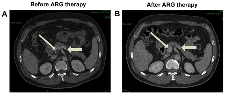
Figure 1.
Contrast-enhanced CT scans of the abdomen in a representative patient with acute superior mesenteric venous thrombosis. (A) Selected axial contrast-enhanced CT image upon admission shows the thrombus located in the superior mesenteric venous trunk, junction of portal (thick arrow) and splenic vein (thin arrow) with bowel edema. (B) CT image at the same level as (A), obtained following argatroban therapy, shows that the thrombus was completely dissolved. CT, computed tomography; ARG, argatroban.

