FIGURE 5.

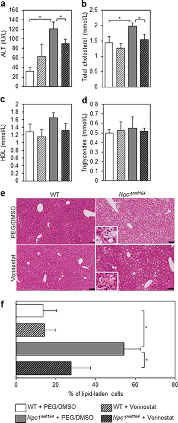
Vorinostat treatment improves liver function in Npc1nmf164 mice. Animals were treated with either vehicle (PEG/DMSO) or 150 mg/kg vorinostat from P21 to P60, at which point serum was harvested from sacrificed animals. Liver function was evaluated via levels of ALT expressed as international units/liter (IU/L; a). Total cholesterol, HDL cholesterol, and triglycerides (b–d) were measured and are expressed as mmol/liter. All data are shown as mean ± S.E. (error bars) for WT + PEG/DMSO (n = 5), WT + vorinostat (n = 5), Npc1nmf164 + PEG/DMSO (n = 4), and Npc1nmf164 + vorinostat (n = 5). *, p < 0.05, Student's t test. e, fixed liver tissue was paraffin-embedded, and 5-μm sections were stained with H&E. Representative images are shown. The prevalence of lipid-laden cells was expressed as a fraction of total cells per field (∼3000) and quantified using SlidePath tissue image analysis (Leica Biosystems, version 2.0) software at ×20 magnification. Data are shown in f as mean ± S.D. (error bars) from 10–15 random fields/animal with 3 animals/treatment group. *, p < 0.001, Student's t test. The inset panels represent a ×80 magnification of the field of view, focusing on the lipid laden cells.
