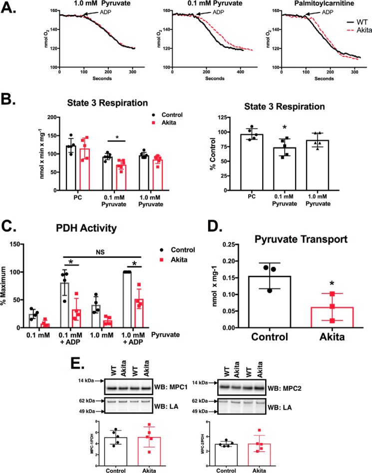
FIGURE 1.
Akita heart mitochondria have impaired pyruvate supported respiration, PDH activity, and pyruvate transport. A, mitochondria were isolated from control and Akita hearts. Respiration was measured by a fiber optic oxygen measurement system with 10 mm malate and either 30 μm PC or the indicated amounts of pyruvate. State 3 was initiated by the addition of 0.5 mm ADP. Representative oxygen traces are shown. B, state 3 respiration rates were quantified and are shown either as specific activities (left) or as the percentage of Akita relative to control rates compared on a day-by-day basis (right; n = 5–6). C, mitochondria were incubated with the indicated amounts of pyruvate for 2.0 min at room temperature. 0.5 mm ADP was added as indicated, and samples were incubated an additional minute. PDH activity was then measured as described under “Experimental Procedures” (n = 4). D, pyruvate uptake was measured in isolated mitochondria as described under “Experimental Procedures” (n = 3). E, MPC1 and MPC2 levels were measured by Western blotting (WB) analysis as described under “Experimental Procedures” (n = 5). The MPC1 and MPC2 Western blots reveal single bands at the expected molecular masses and are cropped for clarity. LA, lipoic acid. Experimental points are from unique mitochondrial preparations, and error bars are the standard deviation. *, p < 0.05, unpaired Student's t test; NS, not significant.

