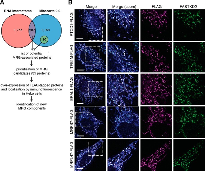
FIGURE 1.
Identification of RNA-binding proteins associated with MRGs. A, schematic representation of the approach used to identify new MRG components. B, representative confocal images of different classes of MRG-associated proteins. HeLa cells were transfected with expression plasmids encoding the FLAG-tagged proteins as indicated. Mitochondria were stained using MitoTracker Deep Red FM (in blue). The cells were immunolabeled with anti-FLAG and anti-FASTKD2 as an MRG-specific marker. White boxes indicate the regions shown at higher magnification. Scale bars are 10 μm.

