
FIGURE 2.
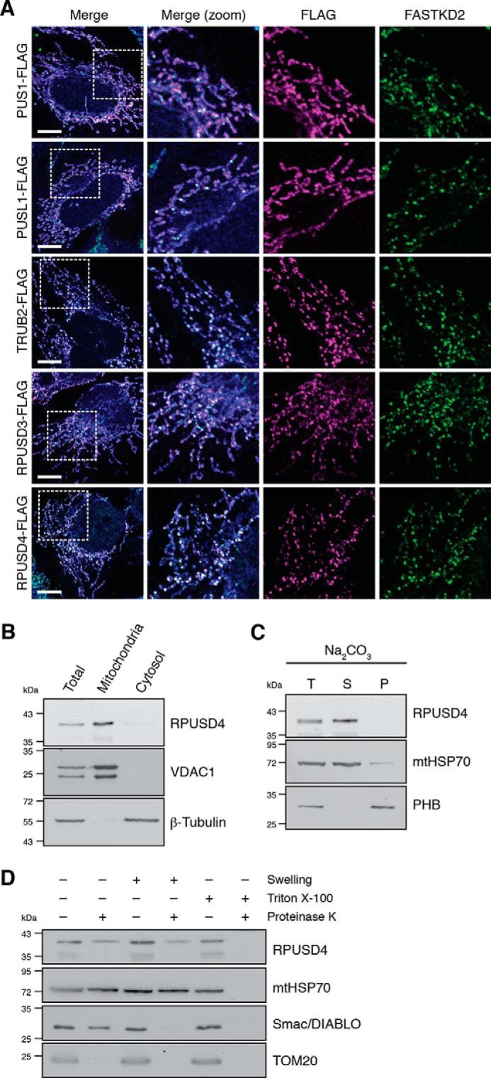
Submitochondrial localization of the mitochondrial pseudouridine synthases. A, confocal analysis of each of the five mitochondrial pseudouridine synthases. 143B cells were transfected with expression plasmids encoding the FLAG-tagged proteins indicated. Mitochondria were stained using MitoTracker Deep Red FM (in blue). The cells were immunolabeled with anti-FLAG and anti-FASTKD2 as an MRG-specific marker. White boxes indicate the regions shown at higher magnification. Scale bars are 10 μm. B, subcellular fractionation of 143B cells and analysis by Western blotting show enrichment for the endogenous RPUSD4 in the mitochondrial fraction. Total cell lysate (T), mitochondrial (M), and cytosolic (C) fractions were assessed for purity using β-tubulin and VDAC1 as cytosolic and mitochondrial markers, respectively. C, immunoblotting analysis of total isolated 143B mitochondria (T) or the supernatant (S) and pellet (P) fractions following alkaline sodium carbonate (Na2CO3) extraction. mtHSP70 and prohibitin (PHB) are markers for the mitochondrial matrix and mitochondrial membranes, respectively. D, immunoblot analysis of isolated 143B mitochondria after proteinase K (PK) accessibility test. Mitochondria were left untreated, swollen to rupture of the outer membrane, or lysed in Triton X-100 prior to treatment with proteinase K. mtHSP70 is a mitochondrial matrix protein, Smac/DIABLO is an intermembrane space protein, and TOM20 is a mitochondrial outer membrane protein.