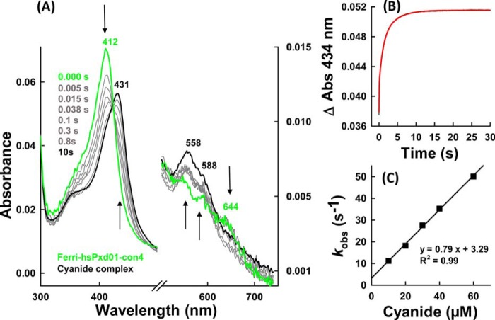
FIGURE 3.
Cyanide binding by ferric hsPxd01-con4. A, spectral changes (arrows) of 1 μm hsPxd01-con4 upon addition of 1 mm sodium cyanide in 100 mm phosphate buffer, pH 7.4. B, time trace and fit (red) of 500 nm hsPxd01-con4 after adding 90 μm cyanide in 100 mm phosphate buffer, pH 7.4. Spectral changes were recorded at 434 nm. C, kobs values for the reaction of 500 nm hsPxd01-con4 reacting with 10–60 μm cyanide in 100 mm phosphate buffer, pH 7.4, plotted against the cyanide concentration for determination of kon, koff, and KD.

