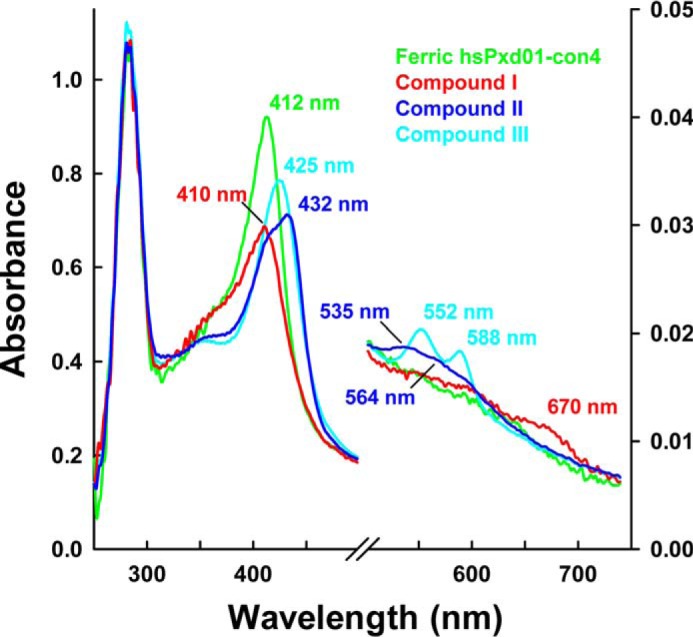
FIGURE 5.

UV-visible spectra of compound I, compound II, and compound III. 1 μm hsPxd01-con4 was reacted with varying concentrations of hydrogen peroxide in 100 mm phosphate buffer, pH 7.4. The ferric form of hsPxd01-con4 is depicted in green, and compound I, compound II, and compound III are shown in red, blue, and cyan, respectively. The characteristic Soret peak maxima and bands in the visible region are illustrated in the same color code. Compound II was formed by the addition of 50 μm hydrogen peroxide, and compound III was generated by adding 1 mm hydrogen peroxide to the ferric protein.
