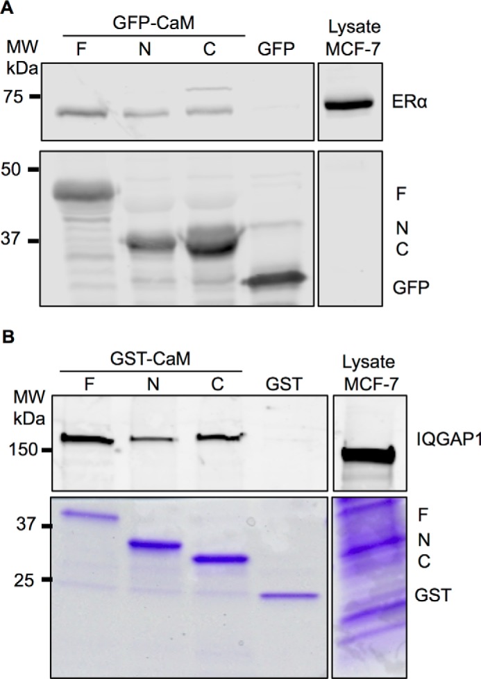
FIGURE 3.

Binding of the CaM constructs to ER-α and IQGAP1. A, HEK-293 cells were transfected with GFP-tagged CaM-F (F), CaM-N (N), or CaM-C (C) or GFP alone (as control). GFP-tagged proteins were isolated with GFP-Trap_A agarose as described under “Experimental Procedures,” then incubated with equal amounts of protein lysate from MCF-7 cells. An aliquot of MCF-7 lysate that was not incubated with GFP-CaM or GFP was processed in parallel (Lysate). Proteins were resolved by SDS-PAGE and Western blots were probed with anti-ER-α (upper panel) and anti-GFP (lower panel) antibodies. Data are representative of 3 independent experiments. B, equal amounts of protein lysate from MCF-7 cells were incubated with GST-tagged CaM-F (F), CaM-N (N), or CaM-C (C) or GST alone (control). Complexes were isolated with glutathione-Sepharose. An aliquot of lysate was processed in parallel (Lysate). Proteins were resolved by SDS-PAGE and the gel was cut at ∼60 kDa. The top part of the gel was transferred to PVDF, whereas the lower part was stained with Coomassie Blue (lower panel). PVDF membranes were probed with anti-IQGAP1 antibodies (upper panel). Data are representative of 2 independent experiments.
