FIGURE 4.

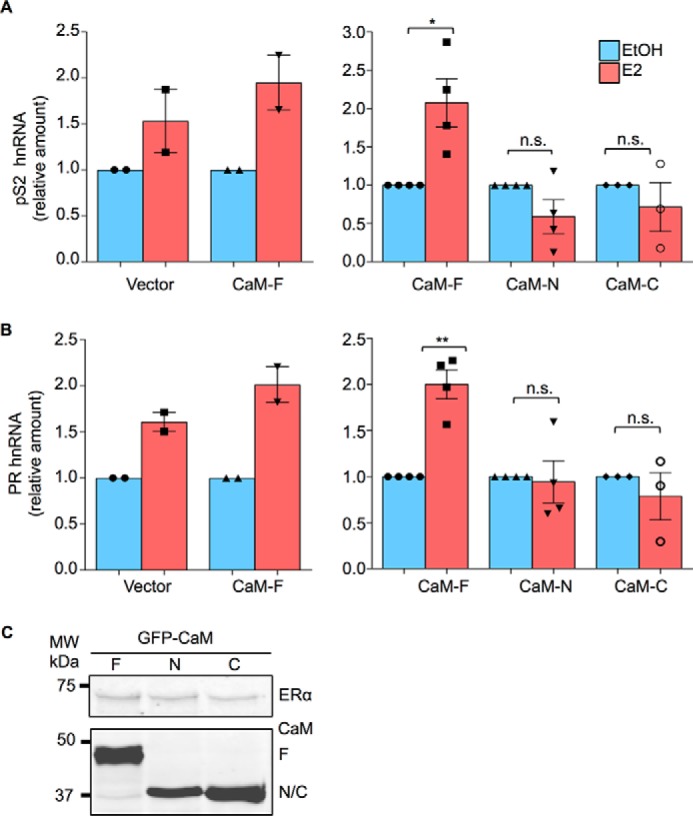
CaM alters ER-α function. A, HEK-293 cells were transiently transfected with both ER-α and GFP-tagged CaM-F, CaM-N, CaM-C, or pEGFP (Vector). Cells were cultured in phenol red-free medium for 24 h, then vehicle (EtOH, blue bars) or 100 nm E2 (red bars) was added to the medium. After incubation for 6 h, total RNA was isolated and quantitative RT-PCR analysis was performed to measure pS2 hnRNA. The amount of RNA in each sample was corrected for β-actin RNA in the same sample. Vehicle-treated cells were set as 1. The data represent the mean ± S.E. (error bars) of two independent experiments for vector and CaM-F (left panel) or three or four independent experiments for CaM-F, CaM-N, and CaM-C (right panel). Each condition was measured in triplicate. B, HEK-293 cells were transiently transfected with both ER-α and either pEGFP (Vector) or the GFP-tagged CaM plasmids. Following cell culture and E2 stimulation, performed as described for panel A, PR hnRNA was measured by quantitative RT-PCR. Samples were analyzed as described for pS2. The data represent the mean ± S.E. (error bars) of two independent experiments for vector and CaM-F (left panel) or three or four independent experiments for CaM-F, CaM-N, and CaM-C (right panel). Each condition was measured in triplicate. *, p < 0.05; **, p < 0.01. C, cells, transfected as outlined above, were lysed and equal amounts of protein lysate were resolved by Western blotting. Blots were probed with antibodies to ER-α (upper panel) and GFP (lower panel). A representative experiment of 2 is shown.
