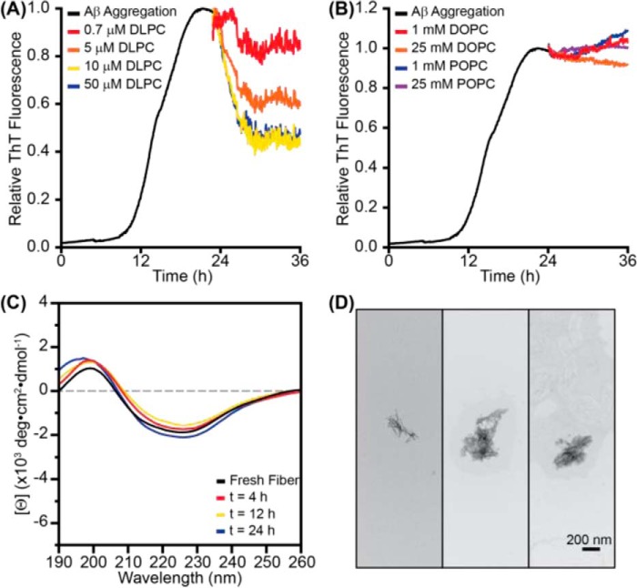
FIGURE 4.
DLPC liposomes remodel preformed Aβ fibrils. A, when 100-nm LUVs of DLPC were added to preformed Aβ fibrils (10 μm monomer concentration; preaggregated for 24 h), ThT (20 μm) fluorescence was decreased in a dose-dependent manner. B, the addition of 100-nm LUVs of DOPC or POPC at much higher concentrations did not impact ThT fluorescence. All ThT curves represent the average of three independent aggregation time courses. C, the CD spectrum of fibrillar Aβ (30 μm) was monitored following the addition of 100-nm LUVs of DLPC (300 μm). D, TEM images of the aggregates induced by incubating preformed Aβ fibrils (30 μm monomer concentration) with 100-nm DLPC LUVs (300 μm lipid).

