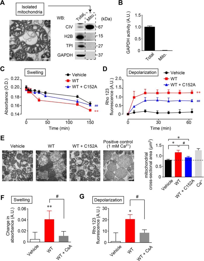
FIGURE 3.
NO-induced aggregates of GAPDH cause mitochondrial dysfunction in vitro. A, transmission electron microscopy of isolated mitochondria (left panel). Scale bar, 400 nm. Western blotting (WB) shows the absence of GAPDH in isolated mitochondria (right panel). B, measurement of GAPDH enzyme activity confirms the absence of GAPDH in isolated mitochondria. C and D, effects of GAPDH aggregation on mitochondrial swelling (C) and depolarization (D). Isolated mitochondria were treated with vehicle (black line and circles), aggregates of WT-GAPDH (0.3 mg/ml, red line and squares), or an aggregate mixture of WT- (0.3 mg/ml) and C152A-GAPDH (0.75 mg/ml, blue line and triangles) for the indicated time periods. Mitochondrial swelling was measured by absorbance at 540 nm. Mitochondrial depolarization was measured by Rho 123 fluorescence. Data are mean ± S.D. (n = 4). **, p < 0.01, relative to vehicle treatment; ##, p < 0.01, relative to treatment with aggregates of WT-GAPDH, Dunnett's test. E, transmission electron microscopy of isolated mitochondria treated with vehicle, aggregates of WT-GAPDH, aggregates derived from a mixture of WT- and C152A-GAPDH, and 1 mm Ca2+ (left panels). Scale bar, 400 nm. A mitochondrial cross-sectional area was determined (right). Data are mean ± S.D. (n = 4). *, p < 0.05, relative to treatment with vehicle; #, p < 0.05, relative to treatment with aggregates of GAPDH, Student's t test. F and G, effect of CsA on GAPDH aggregate-induced mitochondrial swelling and depolarization. Isolated mitochondria were treated with vehicle or aggregates of GAPDH (0.3 mg/ml) or CsA (1 μm) for 2 min prior to treatment with aggregates of GAPDH and incubated for 30 min. Mitochondrial swelling and depolarization were measured as described for C and D above. Data are mean ± S.D. (n = 4). *, p < 0.05, and **, p < 0.01, relative to the treatment with vehicle, Dunnett's test; #, p < 0.05, relative to the treatment with aggregates of GAPDH, Student's t test.

