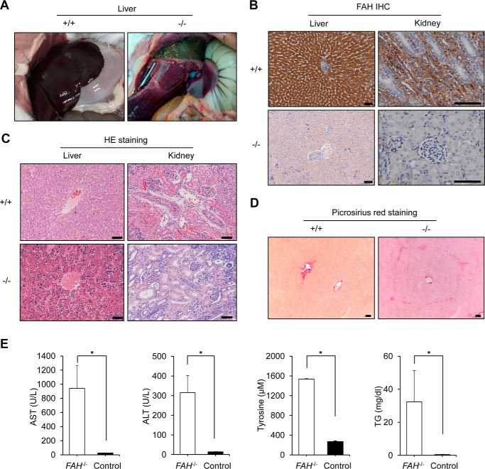
FIGURE 2.
FAH−/− rabbits develop progressive liver failure in the absence of NTBC. A, severe necrosis in the liver of a FAH−/− rabbit (right) belonging to group 3 of NTBC administration (as in Fig. 1D) in contrast to a healthy wild-type rabbit. B, immunohistochemistry (IHC) of liver and kidney sections of a FAH−/− rabbit belonging to group 2 of NTBC administration shows no expression of FAH in contrast to a wild-type rabbit. Scale bars, 50 μm. C, hematoxylin and eosin (HE) staining shows abnormal tissue architecture in liver and kidney sections of the same FAH−/− rabbit in B in contrast to a wild-type rabbit. In the FAH−/− rabbit, diffused hepatocellular injury with dysplastic hepatocytes and tubular epithelial injury of kidney were observed. Scale bars, 50 μm. D, picrosirius red staining shows the existence of mild interstitial fibrosis in the liver of the same FAH−/− rabbit in A but not in the wild-type rabbit. Scale bars, 50 μm. E, serum biochemical parameters indicate liver damage in FAH−/− rabbits compared with a wild-type rabbit. TG stands for triglycerides. Error bars represent S.E. (n = 3 replicate measurements). * corresponds to p < 0.01 according to Student's t test.

