FIGURE 2.
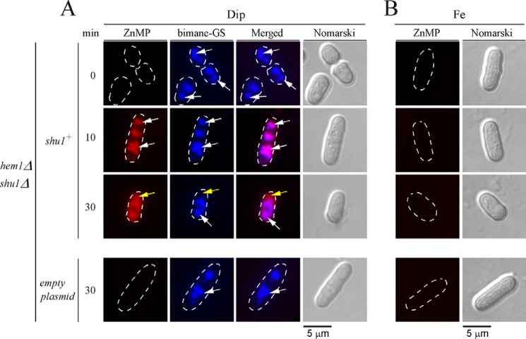
Assimilation of ZnMP is initially detected in vacuoles and then within the cytoplasm. hem1Δ shu1Δ mutant cells expressing Shu1 were precultured in the presence of Dip (50 μm) or FeCl3 (100 μm), and ALA (200 μm). Cells were washed and incubated in ALA-free medium containing Dip (250 μm) or FeCl3 (100 μm) for 6 h. In the final 3 h of treatment, monochlorobimane (100 μm) was added and then ZnMP (2 μm) was added for the indicated times. A hem1Δ shu1Δ double mutant strain in which an empty plasmid was reintegrated (bottom row) was cultured and treated in an identical manner. A, iron-starved (Dip) cells were analyzed by fluorescence microscopy for accumulation of fluorescent ZnMP (far left) and bimane-GS (center left). The merged images are shown in the center right panels. Normarski optics (far right) was used to examine cell morphology. B, as negative controls, iron-treated (Fe) cells are shown because the shu1+ gene is known to be repressed under iron-replete conditions. White arrows indicate examples of vacuoles, whereas yellow arrows depict the cytoplasm where ZnMP was detected after 30 min. Results of microscopy are representative of five independent experiments.

