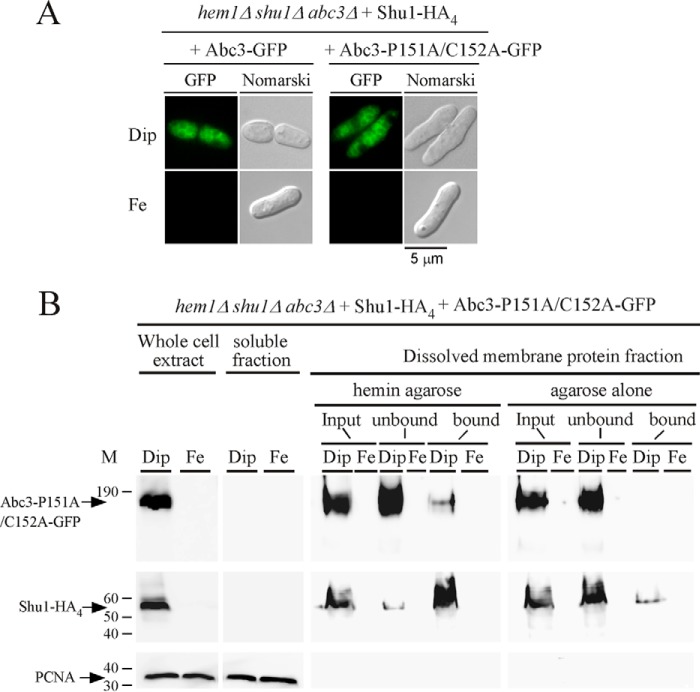
FIGURE 8.
Subcellular localization and hemin-agarose pulldown assays with a mutant version of Abc3. A, fluorescence microscopy of subcellular location of Abc3-GFP or Abc3-P151A/C152A-GFP that was co-expressed with Shu1-HA4 in hem1Δ shu1Δ abc3Δ cells in the presence of Dip (250 μm) or FeCl3 (Fe, 100 μm). Nomarski optics were used to monitor cell morphology. Results of microscopy are representative of five independent experiments. B, whole cell extracts were prepared from cells expressing the mutant version of Abc3 under conditions described above for panel A. Supernatant (soluble proteins) and pellet (membrane proteins) fractions were prepared by ultracentrifugation from whole cell extracts. Triton X-100-solubilized proteins (input) were subjected to hemin pulldown assays using hemin-agarose or agarose. Unbound and bound fractions were analyzed by Western blots. Proteins were revealed using an anti-GFP, anti-HA, or anti-PCNA antibody. The positions of molecular weight standards are indicated on the left. Abc3-GFP, Shu1-HA4, and PCNA are indicated with arrows.

