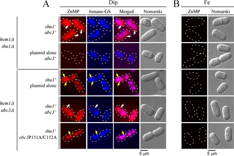
FIGURE 9.
Expression of Abc3-P151A/C152A mutant protein alters ZnMP cellular distribution, leading to its vacuolar accumulation. A, the indicated isogenic yeast strains were cultured in the same manner as described in the legend to Fig. 6. After a 30-min treatment with ZnMP, iron-starved cells were examined by fluorescence microscopy to visualize cellular distribution of fluorescent ZnMP (far left). Vacuoles were detected by bimane-GS staining and cell morphology by Nomarski optics (Nomarski). Merged images of ZnMP and bimane-GS fluorescent signals (center right) are shown next to Nomarski microscope images. B, as negative controls, iron-treated strains are shown because shu1+ and abc3+ genes are known to be repressed under iron-replete conditions. White arrows indicate examples of vacuoles where bimane-GS was sequestered, whereas yellow arrows depict cytoplasmic accumulation of ZnMP. Results of microscopy are representative of five independent experiments.

