
FIGURE 6.
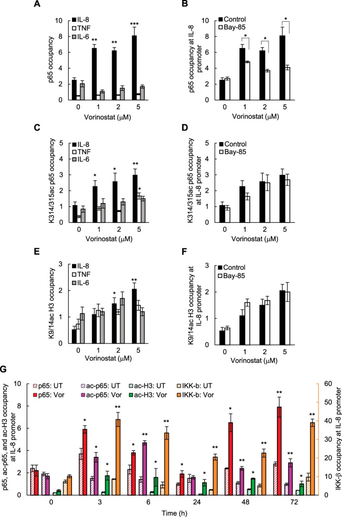
Vorinostat induces p65, Lys-314/315-acetylated p65, and Lys-9/14-acetylated histone H3 recruitment to the IL-8/CXCL8 promoter in ovarian cancer cells. A, ChIP analysis of p65 occupancy at the IL-8, TNFα, and IL-6 promoters quantified by real-time PCR in SKOV3 cells incubated for 48 h with vorinostat. B, ChIP of p65 occupancy at the IL-8 promoter in SKOV3 cells preincubated 12 h with 5 μm Bay 117085 or control DMSO and treated for 48 h with increasing concentrations of vorinostat. C, ChIP of Lys-314/315 ac-p65 occupancy at the IL-8, TNFα, and IL-6 promoters in SKOV3 cells incubated for 48 h with vorinostat. D, ChIP of Lys-314/315 ac-p65 occupancy at the IL-8 promoter in SKOV3 cells preincubated for 12 h with 5 μm Bay 117085 or control DMSO and treated for 48 h with vorinostat. E, ChIP of Lys-9/14 ac-histone H3 occupancy at the IL-8, TNFα, and IL-6 promoters in SKOV3 cells incubated for 48 h with vorinostat. F, ChIP of Lys-9/14 ac-histone H3 occupancy at the IL-8 promoter in SKOV3 cells preincubated for 12 h with 5 μm Bay 117085 or control DMSO and treated for 48 h with vorinostat. G, time course of p65, Lys-314/315 ac-p65, Lys-9/14 ac-histone H3, and IKKβ occupancy at the IL-8/CXCL8 promoter in SKOV3 cells incubated with 1.5 μm vorinostat (Vor) or control DMSO and analyzed by ChIP. The data in A–G are presented as -fold difference in occupancy of the particular protein at the particular locus in comparison with the human IGX1A (SA Biosciences) locus and represent the mean ± S.E. of three experiments. *, p < 0.05; **, p < 0.01; ***, p < 0.001 compared with cells treated with DMSO.