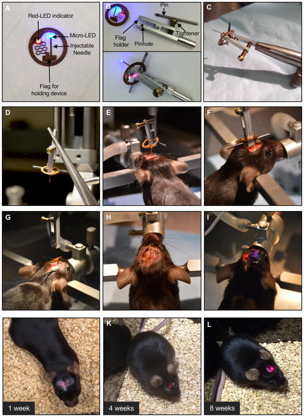
Figure 4. Illustration of surgical procedures for implanting the device and recovered mouse for operation in the deep brain.
A) Representative image of implantable device. B) Images of customized mounting clip and its procedure. C) Image after connecting with stereotaxic arm. D, E, F) Images of the surgical steps for holding and positioning the body of the device, and injecting the needle into the deep brain, respectively. G, H) Images of mouse after releasing of device from stereotaxic arm. I) Wireless operation of implanted device after suturing the skin. J, K, L) Images of recovered mouse after 1, 4 and 8 weeks from surgery, respectively.

