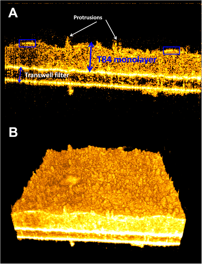
Figure 1. μOCT images of T84 epithelium.

(A) XZ cross-sections. The monolayer (larger blue double sided arrow) is cultured upon a porous Transwell filter (smaller blue double sided arrow). Notable features include apical protrusions (white arrows) and brightly reflective apical surface (blue boxes). (B) 3D rendering showing apical epithelium.
