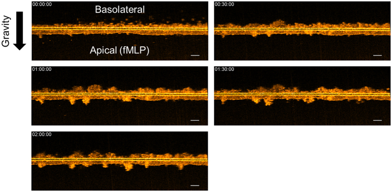
Figure 3. Time-lapse of μOCT cross-sectional XZ images of neutrophils traversing a T84 monolayer in response to fMLP chemoattractant added to the apical compartment.
In the initial frame, neutrophils placed in the basolateral compartment are settling with gravity towards the Transwell membrane. At 30 minutes, neutrophils can be seen entering the epithelium and beginning to protrude into the apical medium. Thereafter, clusters of neutrophils can be seen protruding from the membrane. Scale bars: 50 μm. Full movie data available as Video 4.

