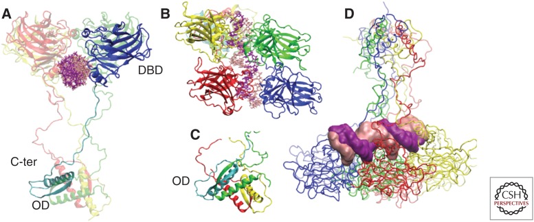
Figure 2.
3D structure of the p53–DNA complex in tetrameric form. (A) Monomer 1 is divided in the DNA-binding domain (DBD) (blue), in interaction with DNA, and carboxy terminal with the OD (oligomerization domain) (cyan). The other three monomers are depicted in transparent mode. (B) Immunoglobulin-like β sandwich architecture of DBD adopted by the four monomers. (C) Tetramerization helices in the carboxy terminal. (D) Projection of the tetramer MD trajectory along eigenvector 1. Specific conformations visited by the tetramer correlate with the DNA deformation measured by the roll and twist parameters (D’Abramo et al. 2015). Therefore, p53 is capable of creating moderate deformities of the DNA, even in the absence of additional molecular partners. C-ter, Carboxy terminal.

