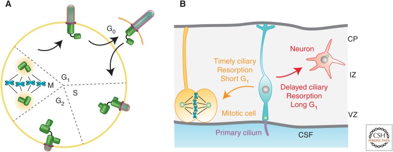
Figure 1.
Ciliary dynamics and cell-cycle progression. (A) Diagram depicts cell-cycle-dependent ciliary assembly and biphasic resorption. (Figure created from data in Bettencourt-Dias and Carvalho-Santos 2008.) (B) Cartoon in which the cell fate choice of a dividing cortical progenitor depends on the time required for ciliary resorption, and, in turn, G1 duration. The primary cilium projecting from the apical endfeet of neural progenitor (in turquoise) faces the ventricle and encounters signal gradient(s) from the cerebral spinal fluid (CSF). VZ, Ventricle zone; IZ, intermediate zone; CP cortical plate.

