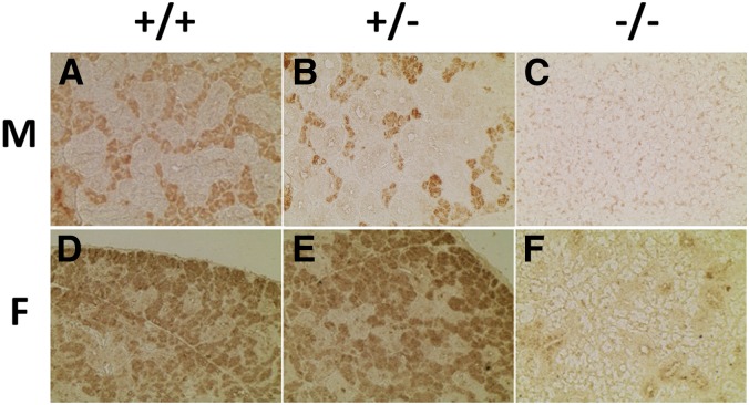
Figure 4.
IHC staining of the submandibular glands of the three genotypes in both sexes. Submandibular gland sections were subjected to IHC staining with anti-ABP antibodies. (A)–(C) are males and (D)–(F) are females, and the three genotypes are shown at the top of the figure. Males have substantially more GCT than females, and that tissue stains negatively (note especially (A) and (B) where stained acinar cells are crowded between GCT). ABP, androgen-binding protein; F, female; GCT, granular convoluted tubules; IHC, immunohistochemical; M, male.

