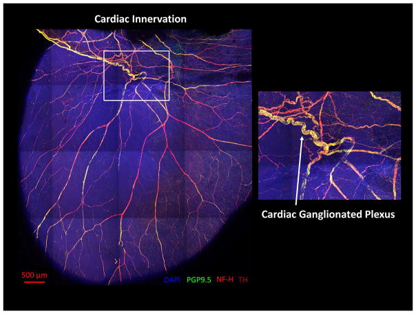
Figure 1.
The “little brain” on the heart. Neural innervation of the murine heart: Confocal microscopic image of a mouse heart stained with antibodies for nuclear marker 4′,6-diamidino-2-phenylindole (DAPI), pan-neuronal marker protein gene product 9.5 (PGP9.5), neurofilament heavy (NF-H), and tyrosine hydroxylase (TH). Mouse heart was cleared using the passive CLARITY technique. Image courtesy of Shivkumar Lab at UCLA and Gradinaru Lab at the California Institute of Technology.

