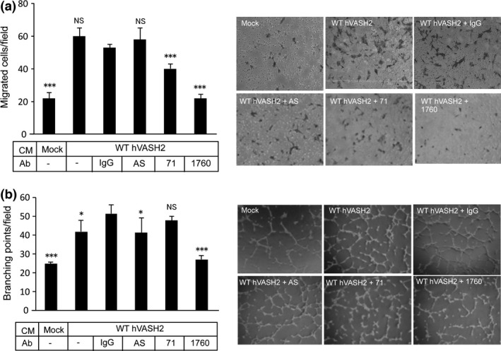
Figure 4.

Selection of neutralizing anti‐hVASH2 monoclonal antibody (mAb). (a) Migration of HUVEC toward WT‐hVASH2 transfectants was examined by Transwell migration assay. Mouse normal IgG (5 μg/mL), antisera or mAb from the indicated clone (5 μg/mL) was added to the lower chamber. The HUVEC that migrated across the membrane were Giemsa‐stained and counted in five random fields at a magnification of ×200. Data are presented as the mean and SD (n = 6). (b) Tube formation by HUVEC on the Matrigel was determined. Mouse normal IgG (5 μg/mL), antisera or mAb from the indicated clone (5 μg/mL) was added to the cultures. Data are presented as the mean and SD (n = 6). Dunnet's test was used for multiple comparisons. *P < 0.05, **P < 0.01, ***P < 0.001 (vs IgG).
