Figure 2.

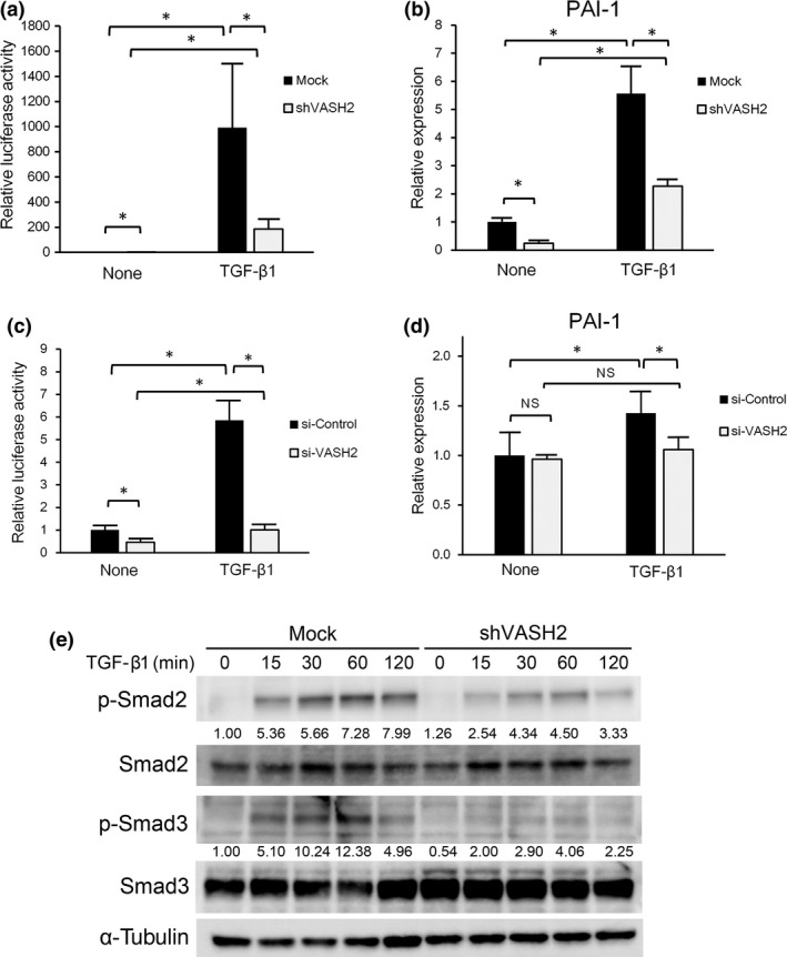
Vasohibin‐2 (VASH2) is required for transforming growth factor‐β (TGF‐β) signaling. (a) SKOV3 cells were cotransfected with the (CAGA)9‐Luc construct and either control siRNA or VASH2 siRNA. Three days after this procedure, cells were treated or not with TGF‐β1 for 24 h, and the (CAGA)9‐Luc reporter activity was then quantified. (b) SKOV3 cells transfected with control siRNA or VASH2 siRNA. Three days after this procedure, cells were treated or not with TGF‐β1 for 24 h. Thereafter, quantitative real‐time RT‐PCR analysis of plasminogen activator inhibitor type 1 (PAI‐1) expression was carried out. Values were normalized to the β‐actin mRNA level. (c) Control mock or shVASH2 transfected cells established from DISS were transfected with the (CAGA)9‐Luc construct. Twenty four hours after this procedure, cells were treated or not with TGF‐β1 for 24 h, and the (CAGA)9‐Luc reporter activity was then quantified. (d) DISS cells transfected with mock or shVASH2 were treated or not with TGF‐β1 for 24 h. Quantitative real‐time RT‐PCR analysis of PAI‐1 expression was then carried out. Values were normalized to the β‐actin mRNA level. (e) DISS cells transfected with mock or shVASH2 were treated with TGF‐β1 for the indicated times then Western blotting for pSmad2, total Smad2, pSmad3, and total Smad3 was undertaken. α‐Tubulin in the cell lysates was used as a loading control. The intensity of each band was determined by densitometry. Values indicate the fold change of pSmad2 and pSmad3 levels normalized to total Smad2 and Smad3, respectively. Mean and SDs are shown (*P < 0.05). N.S., not significant.
