Figure 1.

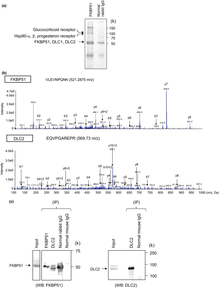
Identification of FKBP51‐interacting proteins. (a) MCF7 cell lysates were immunoprecipitated with normal rabbit IgG or anti‐FKBP51 antibody. The immunoprecipitates were subjected to SDS‐PAGE and visualized by CBB staining. The protein bands specific to FKBP51 were analyzed with LC/MS/MS. In addition to FKBP51, other proteins were identified. (b) MS/MS product ion spectra obtained by nanoflow LC/MS/MS of immunoprecipitated protein complexes from MCF7 cells using an anti‐FKBP51 antibody. (c) MCF7 cell lysates were immunoprecipitated (IP) with rabbit IgG, mouse IgG, anti‐FKBP51, or anti‐DLC2. Total cell lysates and immunoprecipitates were analyzed using anti‐FKBP51 or anti‐DLC2 antibodies.
