
Figure 4.
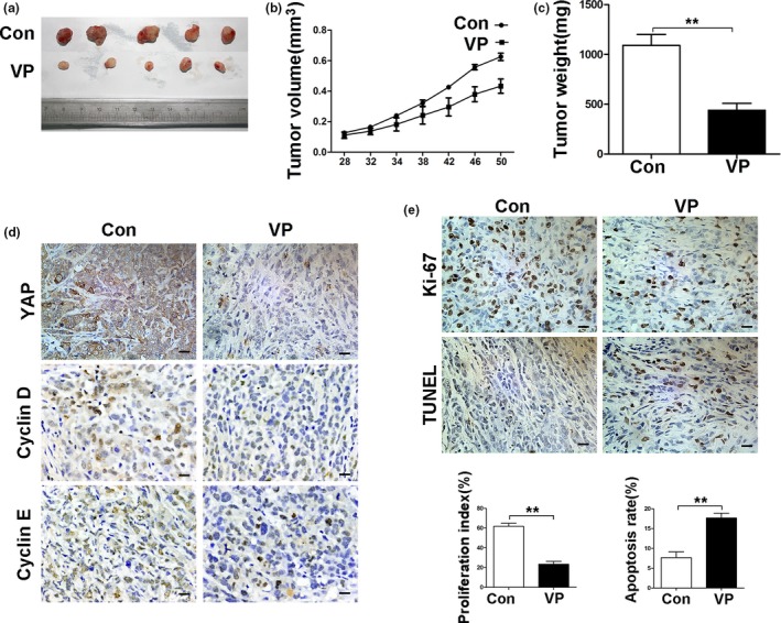
Verteporfin suppresses tumor growth in the PDAC xenograft model. The treatment schedule was shown in Materials and Methods. (a) Tumors were measured and tumor volume calculated at indicated time points. At the end of the experiment, tumors were excised and photographed (b), and weighed (c). (d) Tumoral expression of YAP, cyclinD1 and cyclinE1 was detected by IHC. (e) Tumor sections were stained with an anti‐Ki67 antibody and TUNEL (Magnification× 400, bar = 25 μM). Proliferation and apoptosis indexes were quantified. Data were presented as mean ± SD. *P < 0.05; **P < 0.01; ***P < 0.001.