
Figure 7.
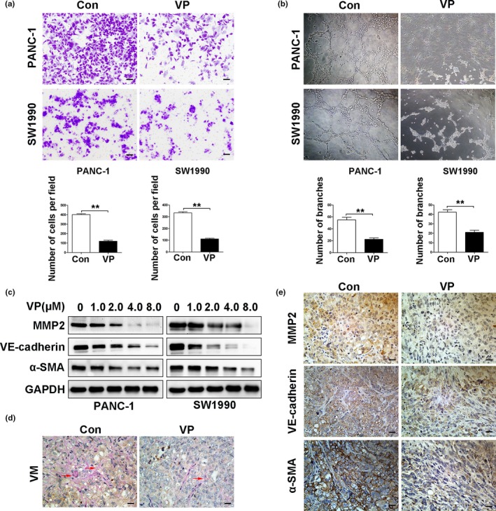
Verteporfin suppresses vasculogenic mimicry in vitro and in vivo. PANC‐1 and SW1990cells were pre‐treated with verteporfin at 4 μM concentration for 24 h. (a) Cell migration was analyzed by transwell assays (Magnification ×100, bar = 100 μM), and (b) VM by tube formation assays (Magnification ×40). The above results were quantified. (c) The expression of MMP2, VE‐cadherin and α‐SMA was detected by Western blot analysis in PANC‐1 and SW1990 cells treated with verteporfin at various concentrations. (d) VM formed in tumors harvested from Figure 4 was detected by staining tumor sections with anti‐CD31 and anti‐PAS antibodies (red arrow, CD31‐negative and PAS positive). (e) The expression of MMP2, VE‐cadherin and α‐SMAin tumors harvested from Figure 4 was detected by IHC (Magnification ×400, bar = 25 μM). Data were presented as mean ± SD. *P < 0.05; **P < 0.01; ***P < 0.001.