Figure 3.

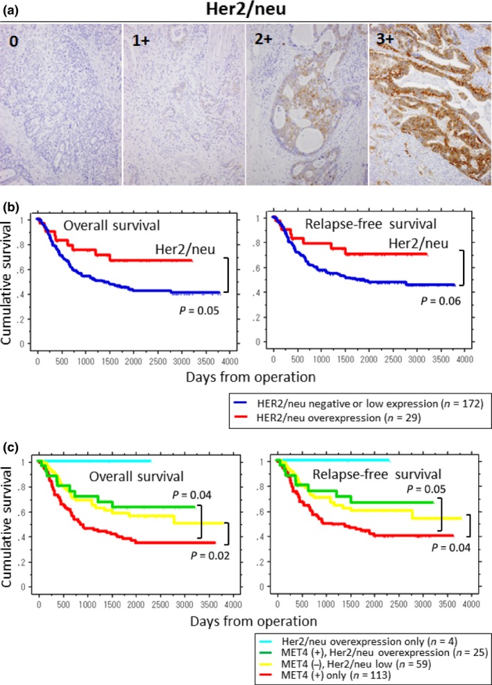
Immunohistochemistry (IHC) staining of Her2/neu and Kaplan–Meier survival plots for overall survival and relapse‐free survival by Her2/neu status. (a) For the evaluation of Her2/neu status in gastric cancers, HercepTest II (DAKO) was used for IHC analysis. Staining levels of 0 and 1+ were dealt with as negative or low expression (without Her2/neu overexpression). Staining levels of 2+ and 3+ were considered as Her2/neu overexpression. (b) Blue lines represent the groups of gastric cancers without Her2/neu overexpression and red lines represent those with Her2/neu overexpression. P‐values of overall survival and relapse‐free survival show 0.05 and 0.06, respectively. (c) Relationship among Her2/neu status, MET4 expression, and prognosis. Patients were divided into four groups according to the expression status of Her2/neu and MET4; cyan: Her2/neu overexpression only (n = 4); green: MET4 (+), Her2/neu overexpression (n = 25); yellow: MET4 (−), Her2/neu low (n = 59); and red: MET4 (+) only (n = 113). The group of MET4 (+) without Her2/neu overexpression (= MET4 (+) only) showed the poorest prognosis.
