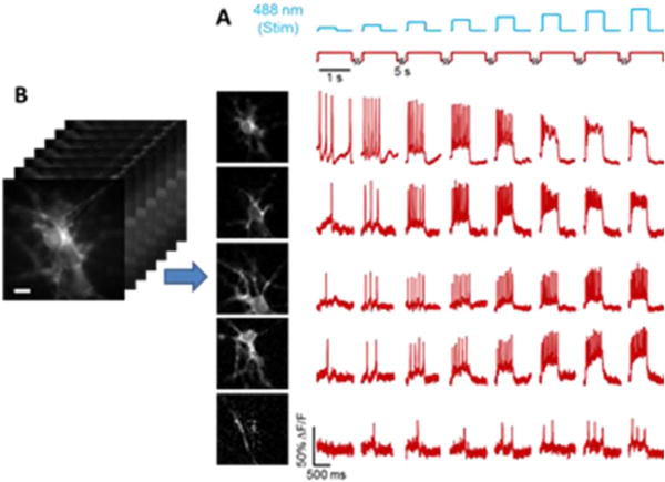
Figure 3.

Activity-based image segmentation. (A) Neurons expressing a channelrhodopsin, CheRiff, and a GEVI, QuasAr2, are stimulated with pulses of blue light and imaged with red excitation and near infrared fluorescence. (B) The cells form a clump, but pixels associated with each cell vary in a unique temporal pattern, allowing a decomposition of the image into a sum of single-cell images, each with an associated firing pattern.
