


Figure 7.
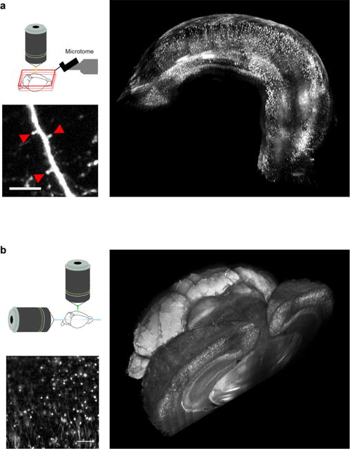
(a) Reconstruction of entire Thy1-GFP-M mouse hippocampus imaged with STP. Magnified insets scale bar = 10 μm. (b) Whole mouse parvalbumin-tdTomato brain tomography obtained with LSFM brain. High resolution insert scale bar = 100 μm. From Costantini et al [60].