


Figure 8.
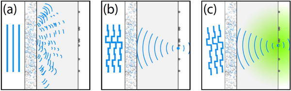
Principle of scattered light fluorescence microscopy (SLFM). (a) A turbid layer blocks a fluorescent structure from sight; all incident light is scattered. (b) The wavefront is shaped with a high-resolution spatial light modulator. For a unique configuration of the modulator, the scattered light interferes constructively at a chosen point; giving rise to a sharp focus. (c) Imaging: the focus follows rotations of the incident beam. The hidden structure is imaged. Reproduced with permission from [66], copyright 2010 OSA Publishing.