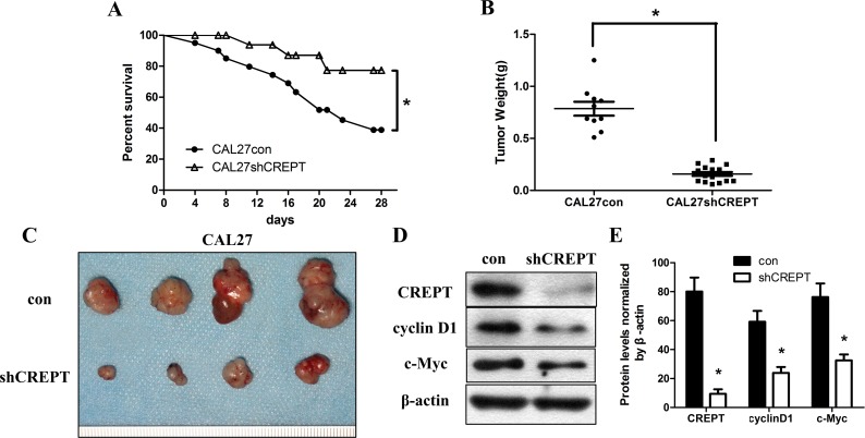
Fig 4. CREPT knocking-down increased survival rate and reduced tumor volume in vivo.
(A) Kaplan–Meier plots of survival analysis of the SCC25 and CAL27 experimental groups compared with their control groups (*p< 0.05, log-rank test). (B) Tumor weights were measured after 28 days(*p< 0.05, Mann-Whitney U test). (C) At 28 days post implantation, tumor xenografts were harvested. Representative images of tumors are shown. (D)CREPT, cyclinD1 and c-Myc proteins in tumor xenografts wereassessed by Western blotting with β-actin as the loading control. The CREPT, cyclin D1 and c-Myc proteins in shCREPT-transfected xenografts were decreased remarkably (*p< 0.05, Mann-Whitney U test).

