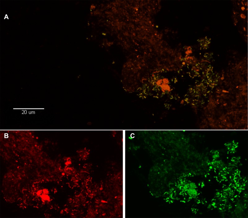
Fig 3. Visualization of P. acnes biofilm in the disc tissue by use of FISH.
A. This color-combined image shows the “pocket” of green fluorescent P. acnes cells (biofilm) near the center right of the image (disc tissue sample #8, Table 2). The presence of P. acnes biofilms in this sample was verified using FISH. B-C. Red fluorescence is the general eubacterial probe (B) and green is the P. acnes probe (C). The B/C image is a zoom of A showing fluorescence from the red and green channels separately. Almost all of the cells in A are emitting both red and green fluorescence indicating that they are P. acnes.

