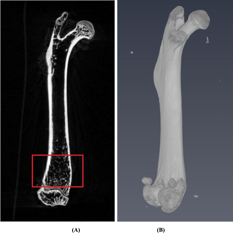
Fig 1.
(A) The growth plate was first located at the distal femur site as a reference plane and delineated a 2 mm distance along the longitudinal direction as shown in the red box for our region of interest. (B) A 3-dimensional surface rendering figure of the whole femur bone which is reconstructed from micro CT images is shown.

