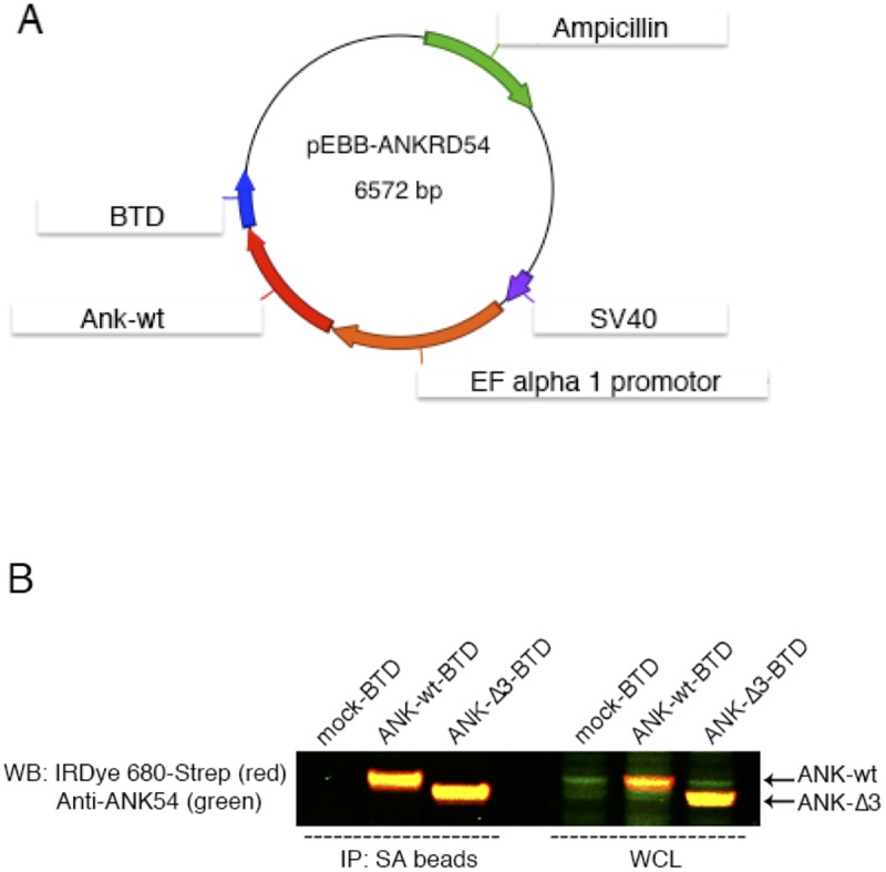
Fig 1. Strategy for protein-analysis and vector validation.

(A) Schematic representation of the pEBB vector containing biotinylation target-domain (BTD) and (B) Simultaneous two-color target analysis of ANKRD54 wt and Δ3 mutant in Cos7 cells. The first three lanes (from left side of the blot) represent immunoprecipitation (IP) with streptavidin (SA) beads and the last three lanes show whole cell lysate (WCL). Anti-ANKRD54 (green) primary antibody recognizes the C-terminus of the protein and anti-IRDye 680-Streptavidin (red) recognizes the BTD domain.
