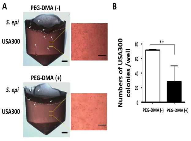
Figure 2.
(A) S. epidermidis counteracts the growth of USA300 in the presence of PEG-DMA in an overlay assay. USA300 bacteria (105 CFU/200 μl; white arrows) were cultured in solid media containing 2% molten agar in rich media with (+)/without (−) 2% PEG-DMA in a 96-well V-bottom PP microtiter plate. S. epidermidis (S. epi) (ATCC12228) bacteria (105 CFU in 20 μl; white arrowheads) were spread on top of the USA300-containing solid media at 37°C for two days. Scale bars=0.1 cm. High magnitude images of USA300 in solid media were displayed. Scale bars=0.5 mm. (B) The numbers of USA300 colonies per well in a 96-well microtiter plate were illustrated as the mean ± standard derivation (SD) of three independent experiments. **P<0.01 (two-tailed t-tests).

