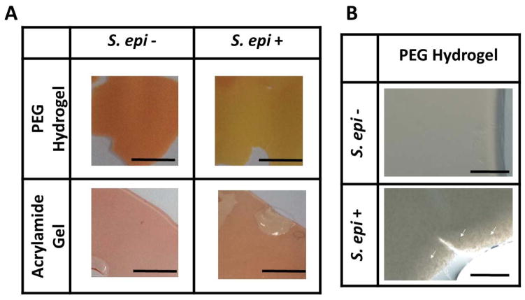
Figure 5.
The fermentation of S. epidermidis in PEG-DMA hydrogels. (A) Hydrogels formed by 10% PEG-DMA (PEG-DMA Hydrogel) or acrylamide (Acrylamide Gel) in phenol red-containing rich media were used to encapsulate the S. epidermidis (2 × 10⁸ CFU/ml; ATCC12228). (B) S. epidermidis indicated with white arrows on the edge of PEG-DMA hydrogels can be visualized in high magnitude images. Hydrogels with (S. epi +) or without (S. epi −) were placed at 37°C for two days. The color change of phenol red from red to yellow in PEG-DMA hydrogels indicated the fermentation of S. epidermidis. Scale bars=0.5 cm; 0.1 cm (high magnitude images).

