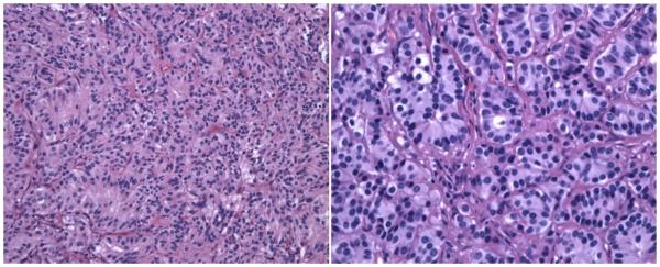
Figure 1.
Hematoxylin and eosin (H&E) staining of the tumor reveals nests of cells (the classic Zellballen pattern) consisting of small ovoid nuclei and eosinophilic cytoplasm separated by thin, fibrovascular septae (H&E × 200 - Figure 1a (left)). Higher power reveals the bland, stippled chromatin pattern and scattered nucleoli (H&E × 400 - Figure 1b (right)).

