
Figure 7. Increased collagen, LOX, and LOXL2 expression predicts poor prognosis among patients with lung adenocarcinoma.
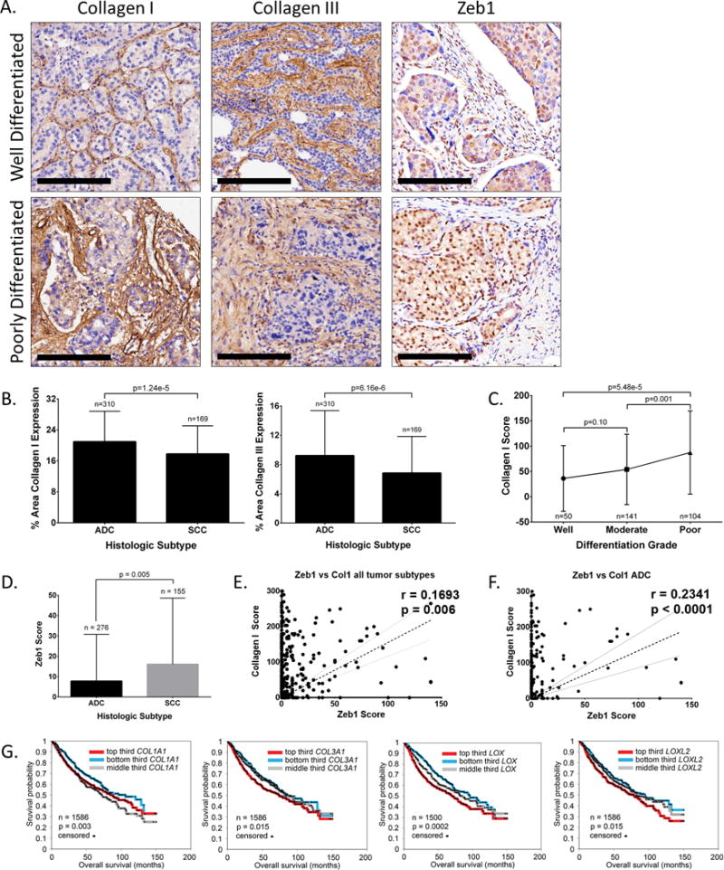
(A) Well differentiated and poorly differentiated human lung adenocarcinoma tissue sections IHC stained for collagen type I, collagen type III, and Zeb1. Scale bars, 200 μm. (B) Percent stromal area of tumor tissues with collagen type I or type III expression in patients with lung adenocarcinoma (ACC) or squamous cell carcinoma (SCC). (C) Average final cytoplasmic H-score of collagen type I expression in lung adenocarcinomas of different grades. (D) Average final nuclear H-score of Zeb1 in tumor cells of ADC or SCC specimens. (E) Cluster plot analysis of Spearman’s rank correlation between Zeb1 and collagen I H-score in both ADC and SCC specimens. (F) Cluster plot analysis of Spearman’s rank correlation between Zeb1 and collagen I H-score in ADC samples. (G) Kaplan-Meier survival analysis by log-rank significance test of COL1A1, COL3A1, LOX, and LOXL2 mRNA expression levels versus overall lung cancer patient survival from a compendium expression dataset of 1,586 lung adenocarcinoma cases. P-values by log-rank test.