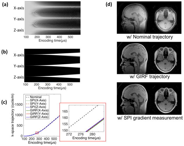
Figure 6.
3D UTE imaging. (a) Log magnitude of k-spaces and (b) normalized images over encoding time in SPI data used for gradient measurement, (c) nominal, GIRF, and SPI measured k-space trajectory, (d) sagittal and axial slice of the image reconstructed with nominal trajectory, GIRF, and SPI measured trajectory. In the images with measured trajectory, no ringing artifact is visible, which is present in the images with nominal trajectory. Note that the coil component is also visible in the images with GIRF and SPI measured trajectory.

