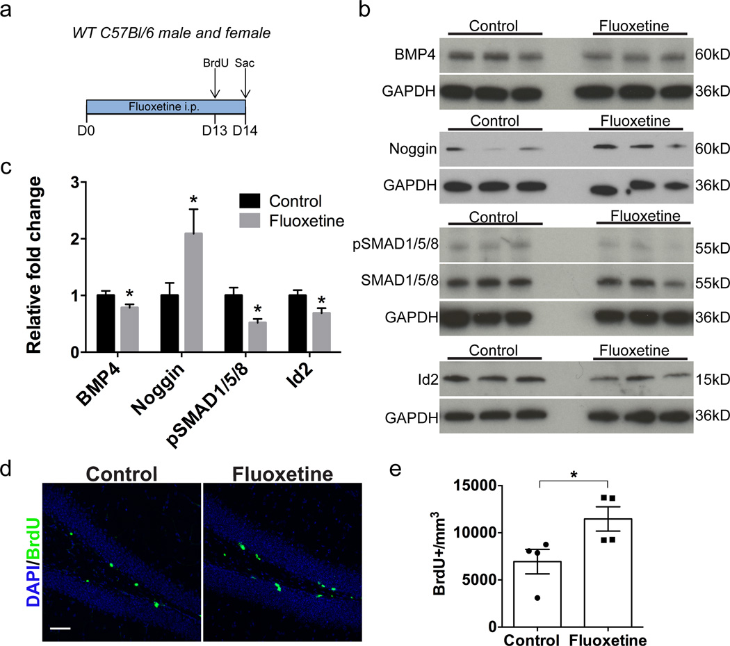
Figure 1. Fluoxetine treatment decreases levels of BMP signaling in the dentate gyrus.
(a) Experimental paradigm: Mice received intraperitoneal fluoxetine or saline once daily for 14 days. On day 13 BrdU was administered to acutely mark the proliferating cell population. On day 14 mice were sacrificed (Sac) and tissue was collected for western blot or immunohistochemical analysis. (b) Western blots of DG tissue show that there is a decrease in expression of the BMP4 ligand and an increase in levels of the BMP inhibitor noggin following fluoxetine treatment. Levels of phospho-Smad1/5/8 and the BMP target gene Id2 are reduced following fluoxetine treatment. GAPDH was used as a loading control. (c) Quantification of western blots by densitometric analysis indicates that there is a decrease in BMP signaling in the dentate gyrus of fluoxetine-treated mice. Values are normalized to loading control and expressed as relative fold compared to saline-treated control mice. n=4–10 per group. (d) Immunostaining for DAPI (blue) and BrdU (green) shows that there is an increase in proliferation in the dentate gyrus following fluoxetine treatment. (e) Quantification of the number of BrdU+ cells per mm3 in the dentate gyrus indicates that there is a 65% increase in the number of BrdU+ cells in fluoxetine-treated mice compared to controls. n=4 per group.
Data are presented as mean±SEM. Unpaired student’s t-test: *p<0.05. Scale bar: 50µm

