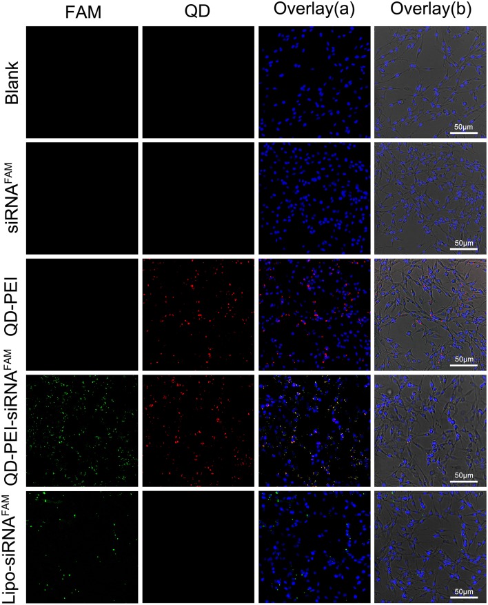
Figure 4.
Laser confocal images of U251 cells treated with different nanoformulations. (I) Blank, (II) free siRNAFAM, (III) QD-PEI, (IV) QD-PEI-siRNAFAM, and (V) Lipo-siRNAFAM for 4 h. Cell nucleus is stained with DAPI (in blue), signals from QD are assigned in red and signals from FAM are assigned in green.

