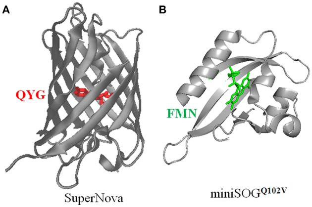
Figure 2.

Three dimensional structures of SuperNova (A), miniSOGQ102V (B), with their respective chromophores highlighted. (A) The full amino acid sequence of SuperNova is obtained from PDB database (3WCK). The three dimensional structure of SuperNova is from PDB in pdb format, input to VMD graphics. The chromophore of Gln65-Tyr66-Gly67 (Takemoto et al., 2013) in SuperNova is highlighted in red. (B) The full amino acid sequence of miniSOGQ102V is from Rodríguez-Pulido et al. (2016). The sequence is put into the protein structure website Swiss-model, three-dimensional model is then obtained after a build-model step. The chromophore FMN in miniSOGQ102V is highlighted in green. Model building similar to Mironova et al. (2013).
