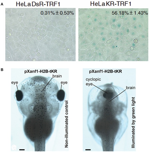
Figure 5.
Photodynamic transformation of cell fate and zebrafish development with KillerRed fusion-expressed with TRF1 at the telomere (KR-TRF1) in HeLa cells or with H2B under the promoter pXanf1 in X. laevis. (A) KillerRed (KR) was fusion-expressed with telomere repeat binding factor 1 (TRF1) to Hela cell telomeres (with the non-photosensitizer DsRed used as control), repeated light exposure (1.5 mW/cm2, 10 min in each passage for 30 passages) induced fastened cell senescence as indicated by enhanced β–galactosidase activity (percentage cells with SA-β-gal staining). The negative control fluorescent protein DsR showed no such effect. (B) KillerRed was fusion-expressed with core histone H2B under the control of forebrain promoter pXanf1, the transgenic embryos were illuminated at the early midneurula stages with green LED (525 nm, 45 mW/cm2, 1 h). Such photodynamic treatment severely retarded the development of the forebrain, sometimes resulted in a completely cyclopic phenotype, similar to phenotypes with suppressed pXanf1 expression (illuminated by green light). The tadpoles developed from transgenic X. laevis embryos not illuminated showed normal phenotype (non-illuminated control). Scale bars in (B) 100 μm. Adapted from Sun et al. (2015) and Serebrovskaya et al. (2011).

