Figure 2.
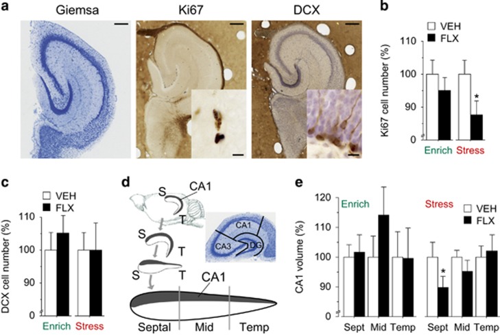
Fluoxetine (FLX) treatment administered in stressful condition leads to a reduction of proliferation and hippocampal volume. (a) Representative sections of histological (Giemsa) and immunohistochemical (Ki67 and doublecortin (DCX) counterstained with hematoxylin) stainings in the mid region of the matrix-embedded, straightened hippocampal dentate gyrus. Scale bar: 250 μm, insert scale bar: 10 μm. (b) Ki67 cell number was not significantly affected by FLX administered in enriched condition, whereas it was significantly decreased in FLX compared with vehicle (VEH) mice when treatment was administered in the stressful condition, *P<0.05 vs VEH group. (c) DCX cell number was not significantly affected by FLX administered in both conditions. (d) Schematic drawing of the straightened hippocampus. Gray part represents CA1. The analysis has been performed independently in the septal, mid and temporal part of the hippocampus, because it has been reported that the effects of SSRIs and the environment are region specific. Boundaries of hippocampal fields are illustrated in the Giemsa-stained section of the mid region of the straightened hippocampus cut perpendicular to the septotemporal axis. S, septal; T, temporal. (e) Analysis of anatomically aligned data of volumetric measurements showed no significant differences between groups in enriched condition. However, septal CA1 volume was significantly reduced in FLX compared with VEH when treatment was administered in stressful condition. *P<0.05. n=8 in all groups. Data shown as mean±s.e.m.

