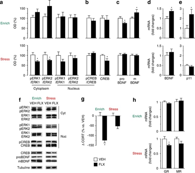
Figure 3.
Fluoxetine (FLX) treatment affects hippocampal signaling pathways and HPA axis activity according to the quality of the environment. (a) No significant treatment effect was found for pERK1/ERK1 and in pERK2/ERK2 ratios in the enriched condition. Whereas both ratios were reduced in the cytoplasmic, but not in the nuclear, fraction in FLX mice in the stressful condition. (b) No treatment effect on CREB phosphorylation was observed in the nuclear enriched fraction in both environmental conditions. No difference in the total hippocampal CREB protein levels was found in the enriched condition, but it was reduced by treatment in the stressful condition. (c) In enriched conditions, reduced proBDNF and increased mBDNF levels were found in FLX mice compared with vehicle (VEH) mice. No difference in the stressful condition. (d and e) Real-time PCR analysis revealed that BDNF and p11 mRNA levels were significantly increased by FLX in the enriched condition but were not affected in the stressful condition. (f) Representative western blottings are shown. (g) Corticosterone levels were significantly reduced by treatment in FLX compared with VEH mice in the enriched but not in the stressful condition. (h) Glucocorticoid receptor (GR) mRNA expression was reduced by treatment in the stressful condition. No difference for GR expression in the enriched condition and mineralocorticoid receptor (MR) expression in both condition was found. MR, mineralocorticoid receptor. Corticosterone analysis: VEH, n= 6; FLX, n=7. For all the other analyses, n=8 in all groups. Data shown as mean±s.e.m. *P<0.05 vs respective VEH group.

