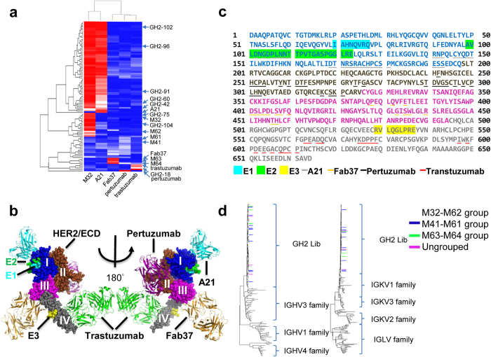
Figure 6. Epitope mapping of anti-HER2/ECD antibodies from GH2 antibody library.
(a) The matrix shows the HER2/ECD binding competitions of the scFvs in S90 against the 4 positive control IgGs, for which the epitopes on HER2/ECD are known from the complex structures determined by x-ray crystallography. (b) Composite complex structure shows the 4 positive control antibodies (PDB codes: 1S78 for pertizumab; 3H3B for A21; 3N85 for Fab37; 1N8Z for trastuzumab and HER2/ECD) binding to HER2/ECD. HER2/ECD is shown in spheres and antibodies are shown in ribbon. Antibody A21, pertuzumab, Fab37 and trastuzumab are colored in cyan, magenta, yellow and green respectively. Domain I ~ IV of HER2/ECD are colored in blue, brown, magenta and gray respectively. Spheres in cyan (E1, domain I), green (E2, domain I) and yellow (E3, domain IV) are the epitopes determined by HDX-MS for GH2 and mouse antibodies; the epitopes of GH2-60, GH2-91, GH2-96, GH2-104, M32 and M62 are designated as E1, the epitopes of GH2-42 and GH2-102 are designated as both E1 and E2; the epitope of M61 is designated as E3. (c) Residue positions of E1, E2, E3, and residue positions of the x-ray structure-determined epitopes (contact surface area > 0) of positive control IgGs are marked on the primary structure of HER2/ECD. The color scheme is the same as in (b). (d) The sequences of variable domains of the S90 scFvs from GH2 library are compared with human germline sequences. The GH2 scFv variants are color-coded at the end of the tree branches based of the assignments of their epitope group (Fig. 5b): scFv variants in the M41-M61 epitope group is highlighted in blue, M63-M64 epitope group in green, and M32-M62 epitope group is not colored; scFvs with ungroup epitopes are highlighted in magenta. The human germline sequences were attained from the IMGT database. The plot shows that these epitope groups are not correlated with sequence relationships.

