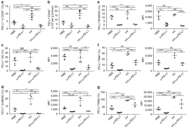
Figure 5. The α-PD-L1 treatment reduces the PD-L1+ cells in TME.
B6 mice were intraperitoneally inoculated with 5 × 10⁵ MC38-luc cancer cells and treated with VV and/or α-PD-L1 as described. Tumour-bearing mice were killed at day 5 post first treatment and primary tumours were collected and analysed to determine the PD-L1+ CD45− cells (a,b), PD-L1+ DC (defined as CD45+ CD11b+CD11c+Ly6G−PD-L1+) (c), PD-L1+G-MDSC (defined as CD45+CD11c−CD11b+Ly6G+Ly6clowPD-L1+) (d), PD-L1+ M-MDSC (defined as CD45+CD11c−CD11b+Ly6G−Ly6chiPD-L1+) (e), PD-L1+ TAM1 (defined as CD45+CD11c−CD11b+Ly6G−F4/80+CD206−PD-L1+) (f), PD-L1+ TAM2 (defined as CD45+CD11c−CD11b+Ly6G−F4/80+CD206+PD-L1+) (g). Of note, the anti-PD-L1 antibody clone 10F.9G2 was used for therapy while clone MHI5 was used for subsequent analysis. Data were analysed using Student's t-test (*P<0.05; **P<0.01; ***P<0.001; ****P<0.0001).

