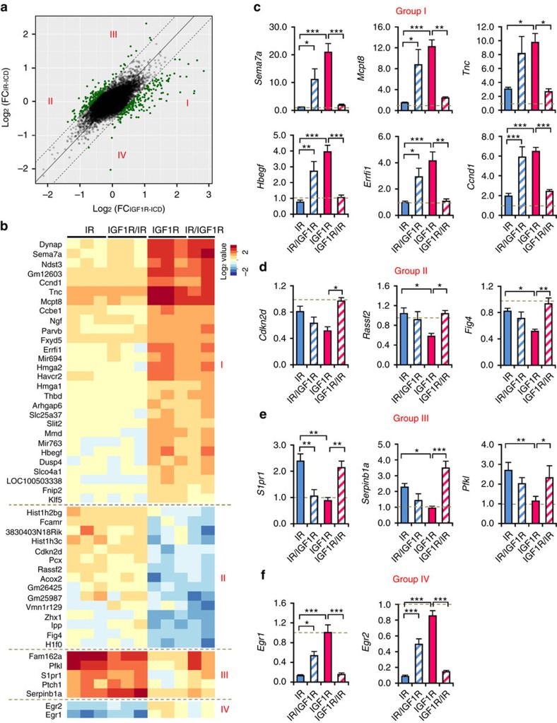
Figure 5. Differential roles of IR-ICD and IGF1R-ICD in regulating gene expression.
(a) Scattered plot showing distribution of log fold change of each probeset's expression after stimulation in cells expressing receptors with IR-ICD (y axis) versus that in IGF1R-ICD (x axis). Each dot represents the mean of all the clones expressing the receptors with the same ICD. The highlighted dots represent the probe sets with fold-change difference between IR-ICD and IGF1R-ICD over 50%. (b) Heat map of top significant genes in a classifying into four groups. (c,d) Fold changes of representative genes highly induced (c) or suppressed (d) by cells expressing receptors with IGF1R-ICD were confirmed by qPCR using TBP as a standard. (e,f) Fold changes of expression in response to stimulation for representative genes preferentially induced (e) or suppressed (f) by cells expressing receptors with IR-ICD were confirmed by qPCR using TBP as a standard. Data are mean±s.e.m. (*P<0.05; **P<0.01; ***P<0.001. One-way ANOVA followed by Newman–Keuls post-hoc analysis, n=6).

